Advice on a fungal ID
- Thread starter rich59
- Start date
You are using an out of date browser. It may not display this or other websites correctly.
You should upgrade or use an alternative browser.
You should upgrade or use an alternative browser.
Hi Rich, Do you know what type of wood it was on? looking at it i think it could be Beech Jellydisc Neobulgaria pura.
I don't know what the wood was. It could have been beech, the bark was smooth and gray. On similar logs in the same area was the ear fungus (usually on elder) and cramp balls (usually on ash).
I looked at some pictures of your suggestion. Seemed a bit translucent and not so cup like as my specimen.
I looked at some pictures of your suggestion. Seemed a bit translucent and not so cup like as my specimen.
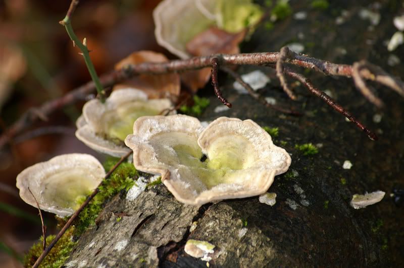
rich59 said:This chap I found today. It is growing on dead wood, not on the ground. That confused me as otherwise it could have been the orange peel fungus. Any thoughts?
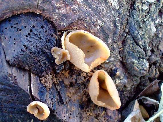
looks like one of the peziza fungi... repanda or cerea maybe?
roger philips guide: p 269

xylaria
Bushcrafter (boy, I've got a lot to say!)
We are having a problems IDing this one. There is alot a candates which are very simerlar being put forward. I think we may have to look at this one in a differant manner.
The fundamantal differances used for the identification of ascomyctes differ from that of the gill fungi. The structure of the fungus, and the look of the spores are alot more important. If think abot horses hoof fungus the cross section view is easyest way of being sure of what you have. With smaller fungi a hand lens is nearly always needed to get same type of view of the cross section. I have collected fungi for years without needing a microscope. For mycrophagy a microscope feels like over kill to me, but even I admit sometimes they are needed for certain types of non-edible fungi. Cup fungi are one of those groups.
Under a standard microscope (not oil emersion) a thin slice of the cross section of fruit body is taken and dyed with melzar reagent. Below 500X mag the spore baring stuctures should be visable. There will be various layers of cells that form the outside of the fruit body. The important bit to look at is the spore baring inner area. The acsi (where the spores form) and the paraphyses (sterile) should be projecting out of the lower layers like a grass lawn. They are 250microns long so are much bigger than spores. Basically the paraphyses come in a variety of shapes: branched, forked, hooked, clubed, and straight. These may visable without dye, I dont know I havent looked at anything other than spores for good many years.
Humaria hemisphaerica has hairs on the outside the are awl-shaped when veiwed with a hand lens.
Neobulgaria pura is more flat and jelly like.
I personally I feel it may be a peziza. Considering it is growing in a normal place (not a fire site or an office carpet") ) it is really hard it say what it is.
) it is really hard it say what it is.
Crepidotus spp are gilled. The white braket fungi is not.
Coriolus hirsutus sounds good I don't know of anything better that fits.
The fundamantal differances used for the identification of ascomyctes differ from that of the gill fungi. The structure of the fungus, and the look of the spores are alot more important. If think abot horses hoof fungus the cross section view is easyest way of being sure of what you have. With smaller fungi a hand lens is nearly always needed to get same type of view of the cross section. I have collected fungi for years without needing a microscope. For mycrophagy a microscope feels like over kill to me, but even I admit sometimes they are needed for certain types of non-edible fungi. Cup fungi are one of those groups.
Under a standard microscope (not oil emersion) a thin slice of the cross section of fruit body is taken and dyed with melzar reagent. Below 500X mag the spore baring stuctures should be visable. There will be various layers of cells that form the outside of the fruit body. The important bit to look at is the spore baring inner area. The acsi (where the spores form) and the paraphyses (sterile) should be projecting out of the lower layers like a grass lawn. They are 250microns long so are much bigger than spores. Basically the paraphyses come in a variety of shapes: branched, forked, hooked, clubed, and straight. These may visable without dye, I dont know I havent looked at anything other than spores for good many years.
Humaria hemisphaerica has hairs on the outside the are awl-shaped when veiwed with a hand lens.
Neobulgaria pura is more flat and jelly like.
I personally I feel it may be a peziza. Considering it is growing in a normal place (not a fire site or an office carpet
) it is really hard it say what it is. Crepidotus spp are gilled. The white braket fungi is not.
Coriolus hirsutus sounds good I don't know of anything better that fits.
xylaria said:We are having a problems IDing this one. There is alot a candates which are very simerlar being put forward. I think we may have to look at this one in a differant manner.
The fundamantal differances used for the identification of ascomyctes differ from that of the gill fungi. The structure of the fungus, and the look of the spores are alot more important. If think abot horses hoof fungus the cross section view is easyest way of being sure of what you have. With smaller fungi a hand lens is nearly always needed to get same type of view of the cross section. I have collected fungi for years without needing a microscope. For mycrophagy a microscope feels like over kill to me, but even I admit sometimes they are needed for certain types of non-edible fungi. Cup fungi are one of those groups.
Under a standard microscope (not oil emersion) a thin slice of the cross section of fruit body is taken and dyed with melzar reagent. Below 500X mag the spore baring stuctures should be visable. There will be various layers of cells that form the outside of the fruit body. The important bit to look at is the spore baring inner area. The acsi (where the spores form) and the paraphyses (sterile) should be projecting out of the lower layers like a grass lawn. They are 250microns long so are much bigger than spores. Basically the paraphyses come in a variety of shapes: branched, forked, hooked, clubed, and straight. These may visable without dye, I dont know I havent looked at anything other than spores for good many years.
Humaria hemisphaerica has hairs on the outside the are awl-shaped when veiwed with a hand lens.
Neobulgaria pura is more flat and jelly like.
I personally I feel it may be a peziza. Considering it is growing in a normal place (not a fire site or an office carpet
Crepidotus spp are gilled. The white braket fungi is not.
Coriolus hirsutus sounds good I don't know of anything better that fits.
I agree with the Peziza, it looks a tad transparent for Otidea alutaccea which would infer it growing on soil in those cracks! An iodine test should distinguish, Peziza will blue at the edges Otidea won't.
I went back to the site and took a close look to see what it was growing from - soil or wood. Definitely from the wood. There was no soil. There were several samples of it on 2 logs. One sample was close to a crop of something like turkey tailPipistrelle said:I agree with the Peziza, it looks a tad transparent for Otidea alutaccea which would infer it growing on soil in those cracks! An iodine test should distinguish, Peziza will blue at the edges Otidea won't.
I think I read that some peziza can grow both on wood and on the ground.
I have a small microscope but it is not good enough to resolve spores apart from vague specs. Xylara - could you recommend what standards of magnification and resolution I would need to differentiate spores well?
xylaria
Bushcrafter (boy, I've got a lot to say!)
Thanks piperstrelle I didn't know about the iodine test, it sounds really simple
Rich59 The standard student microscope you discribed in previous thread that you have should be adiquit to view the parphysis (plural? ). The only way I have seen the features on a spore is a 1000x bifocal in uni. These are normally very costly and most fungi nuts tend to get by with a normal microscope viewing what you can view. You do get the hardend kit junkys with mycologists who would have an eletron mircoscope if they could fit it in the shed. 
Remember to calculate the strenght of magnification is to muliply the eye piece by the lens. If I can recall you had 10X at eye and 20x at lens so that is a 200x mag.
Rich59 The standard student microscope you discribed in previous thread that you have should be adiquit to view the parphysis (plural?
). The only way I have seen the features on a spore is a 1000x bifocal in uni. These are normally very costly and most fungi nuts tend to get by with a normal microscope viewing what you can view. You do get the hardend kit junkys with mycologists who would have an eletron mircoscope if they could fit it in the shed. Remember to calculate the strenght of magnification is to muliply the eye piece by the lens. If I can recall you had 10X at eye and 20x at lens so that is a 200x mag.
Similar threads
- Replies
- 2
- Views
- 227
- Replies
- 10
- Views
- 263
- Replies
- 7
- Views
- 245